Patient information and CT scan

What examinations are performed on the CT scan?
What are the preparations and what is the aftercare?
The different examinations were placed here according to anatomical location:
From the head to the feet.
Navigation : Head – Neck – Vertebral column – Thorax – Abdomen/pelvis – Skeleton
CT scan of the head
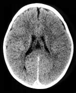
A CT scan of the brain can be performed with or without contrast administration. Which method is used depends on the indication. In some cases, it may be appropriate to perform the examination first without and then with contrast to obtain a clearer picture in the patient’s pathology.
In cases of trauma, the radiologist may also interpret the images taken in a window where bone structures are more clearly visible.
If contrast will be administered, it is necessary to be sober for the examination. For this, it is best not to eat or drink for at least four hours before the examination.
Back to navigation
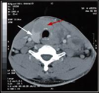
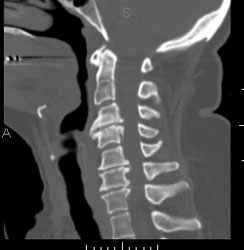
CT Scan of the neck
The purpose of a scan of the neck may be to detect a defect at the level of the blood vessels of the neck, or an abnormal occurrence of a certain structure in the neck, e.g. an enlarged thyroid gland.
A CT scan of the neck is usually performed with contrast.
Exceptions to this are examinations of the thyroid gland.
If contrast is given, the patient must be kept fasting for at least four hours before the examination.
CT scan of the vertebral column
A ct scan of the vertebral column is generally requested to assess lesions at the level of the vertebrae, or to check the permeability of the spinal canal. Think here of detecting a disc herniation, or a traumatic injury of a vertebra.
Basically requires no specific preparation.
The patient lies supine on the examination table and must remain so until after the examination. It is important here that the patient does not change position again.
Contrast is administered here in some cases, but this is not a rule.
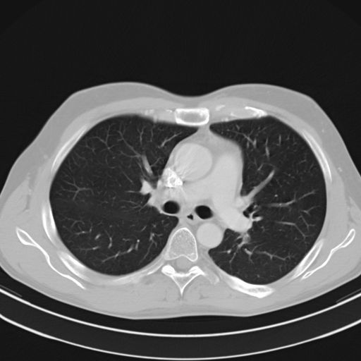
CT scan of the thorax
Here an image of the structures of the chest is formed. The lungs, mediastinum as well as the blood vessels and heart are assessed. During the examination, the patient will be asked to hold their breath. This is to get an even follow-up of the images. Also, it is obviously important not to move during the examination. However, due to the ever-increasing speed of the devices, this is not a difficult task, if you think that a complete lung scan takes about thirty seconds or so. However, it is possible that a second recording will be taken for the sake of completeness, so staying down until you are told it is over is the message.
Depending on the indication, a decision is made to administer contrast or not.
Again, it is then necessary to keep the patient sober at least four hours before the examination.
Back to navigation
CT scan of the abdomen or small pelvis
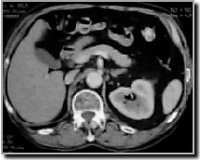
A CT scan of the abdomen or small pelvis is taken to assess any problems at the level of the intestines, liver, kidneys, spleen, etc…. assessment.
Again, it is very important that the patient not move during the examination. You will be asked to hold your breath several times. This is also important to ensure good continuity of images.
Except when the examination is done to get an image of blood vessels and bone structures, contrast is generally always given for drinking. Intravenous contrast is administered except for bone studies.
This is where good preparation is then needed.
A cleansing laxative or oral laxative will be given the day before the examination.
The day of the examination, the patient should drink one liter of contrast medium over an hour’s time. This is to fill the intestines as much as possible. This way, a clear image of the intestines can be formed. A lavage with contrast is also sometimes necessary. This is to fill the last parts of the intestine.
(Note: In some centers, instead of a lavage, they give a bottle of diluted contrast as early as the day before. However, this gives a less clear picture of the last bowel lesions).
Back to navigation
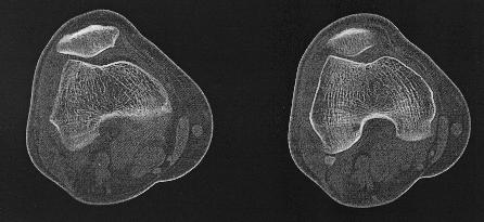
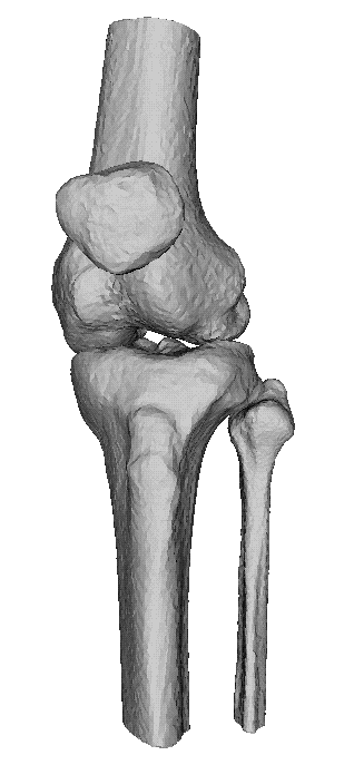
CT of the skeleton
A Ct-scan of the skeleton is performed when further evaluation is needed of an injury seen on a classic photograph. Think of fractures, luxations, etc., but also metastases, osteoarthritis,…For example, it may be that an injury does not appear clearly enough on the photos and therefore there is doubt. But it may also be that a CT scan is necessary to obtain a clear picture of an extensive fracture, to determine further treatment.
Thanks to modern computer techniques, it is not only possible to make axial images. (read slices across the body) But also to reconstruct those images into a 3D model. This often gives a clear picture of the bone and its injury.
Back to navigation