


To give a picture of the evolution of angiography, we must follow two parallel evolutions. The development of X-ray techniques, necessary to obtain the visualization of blood vessels, as well as the emergence of catheters and related techniques.
We find the first forms of catheterization as early as 3000 B.C. where the Egyptians performed bladder catheterizations with metal tubes. As early as 4000 B.C., hollow straws were used to study hearts of corpses.
In 1711, Hales inserted a catheter made of glass and the trachea of a goose into the heart of a horse.
In 1844, Claude Bernard managed to insert a mercury probe into the carotid artery of a horse and push it further up into the left anterior chamber, this to measure the pressure in the blood. Over the next forty years, he further developed and improved this technique, and then applied it to other animals. His pioneering work led to the forms of catheterization used today.
In 1896, the first image of blood vessels was made by using rays and contrast media. lime was then used that was injected into the blood vessels of an amputated hand of a corpse. This was done by Hascheck and Lindenthal.
The first catheterizations performed on humans were mainly those of Fritz Bleichroeder, E. Unger and W. Loeb in 1912. They were the first to insert catheters into humans, but without using X-rays.
Also, the chemotherapy that emerged during that period required the use of catheters. This was to administer the medication directly into the blood. Fritz Bleichroeder inserted catheters into the arteries of dogs and left them in place for a few hours, noting that there were no complications, not to mention the formation of clots.
On June 26, 1926, Egaz Moniz obtained on his ninth attempt, a successful angiogram of the head in a living patient. This is therefore the first documented angiographic examination in living people. For this, iodine was injected into the patient’s carotid.
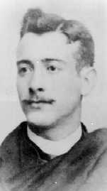
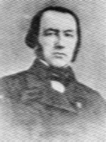
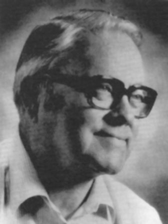
Werner Forssmann Ziedses Des Plantes
In 1929, Germany’s Werner Forssmann experimented with urological catheters on human corpses. He discovered that advancing and catheter from the arm to the right ventricle through the veins was very easy. He then went so far as to insert such a catheter into his own arm and advance it to his ventricle under fluoroscopy guidance, using a mirror to follow himself. This made him the first person to have a cardiac catheterization. For developing that technique and taking it further, he was awarded the Nobel Prize in 1956.
In 1934, a young student, Ziedses Des Plantes, came up with two new techniques in his thesis that would greatly influence radiology: tomography and subtraction. For the latter, he superimposed a negative image without contrast addition on a positive image with contrast addition, thus obtaining a subtraction image showing only the contrast. This technique was used for about fifty years before the computer took over the task of subtraction.
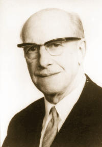
In 1953, Mr. Sven Ivar Seldinger developed a technique for smoothing both the left and right heart. This technique was widely adopted and applied by other researchers and is now the standard method of puncturing blood vessels for catheterization.
Between 1940 and 1950, various techniques were developed to examine blood vessels, including the injection of contrast media.
In 1964, Dr. Charles T. Dotter discovered the concept of repairing blood vessels by working inside the blood vessel itself, or transluminal angioplasty. And in 1977, the first balloon dilatation was performed by Drs. Andreas Gruentzig, Myler and Hanna at San-Fransico, this for the first time in a cath lab and only under fluoroscopic guidance. This is important since balloon dilations were previously done under anesthesia in an operating room….
In 1967, Kurt Amplatz, G. Formanek, P. Stranger and W. Wilson developed catheters with special preformed bends to be used in catheterization when puncturing the arteria femoralis.
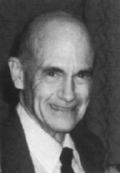
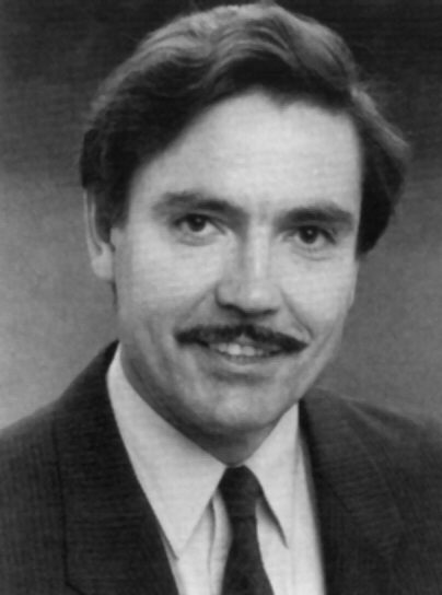
Sven Seldinger Charles Dotter
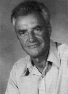
Andreas Gruenzig Kurt Amplatz
In the following years, several innovations were made that would determine the future of angiography.
Inventing the guide wires and “steerable” catheters and a number of systems that further advanced angioplasty.
One is now able to solve many of the blood vessel problems by taking an access route along the blood vessels themselves.
These include embolization (stopping bleeding or perfusion), dilation (widening of clogged vessels), stenting (applying support systems for the blood vessel wall to prevent clogging),to removing plaques and deposits on the vessel walls,…
The use of laser did not become the hoped-for success, but other systems were accepted and used worldwide.
Such as rotary atherectomy systems (rotablator) and intravascular ultrasound.
2002 marked the 25th anniversary of angioplasty in “awake” patients.
i.e., without anesthesia.
As the MRI and CT scan more a more displace classical angiography for diagnostic purposes, angiography is more appropriate for direct treatment.
Angioplasty is still on the rise.
Studies are currently underway to apply angiography under MRI guidance.
Although that technology is still in its infancy, it already appears that it will be used more in the future.
In any case, it opens up more possibilities that are safer for both the patient and the researcher, if only because X-rays are no longer needed.