


Principle
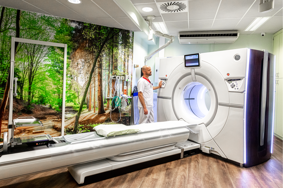
Usually, one uses a system in which the X-ray tube is mounted on an annular structure that lies around the patient and on which the tube can rotate all the way around. On the opposite side of the tube are detectors on the ring that collect the radiation after penetration of the patient.
The patient lies on a tabletop that brings the region to be examined into the center of the gantry. This sheet can be shifted to the millimeter precisely so that one can determine very precisely which region to study.
To create images, the rays attenuated by the various tissues are measured by the detectors. These have the property of converting the incident radiation into a proportional electrical signal. By rotating the tube around the patient, images can be taken in different directions. The computer collects all the data and can thus reconstruct an image, called a coupe or cut.
The image one obtains is composed of different shades of gray or densities. Each tissue has its own density and therefore has the ability to attenuate radiation more or less depending on its density. Densities were determined in Houndsfield Units (H.U.).
| Bot | > 250 H.U. (white, very high density) |
| Liver | 65 H.U. |
| Water | 0 |
| Fat | – 65 H.U. |
| Air | – 1000 H.U. (black, very low density |
Spiral CT
Since the 1990s, a new technique called spiral or volume CT has been used. Unlike classical CT where the patient was moved over a certain distance each time before taking an image, here the patient is moved at a constant speed under continuous rotation of the tube.
Due to the continuous movement of the patient and the rotation of the tube around it, a spiral structure is obtained.
An entire volume is scanned at once.
The advantages are:
 | The investigation ends more quickly |
| More data are obtained, allowing reconstruction in other planes (sagital, axial) |
| It gives the ability to create 3D models |